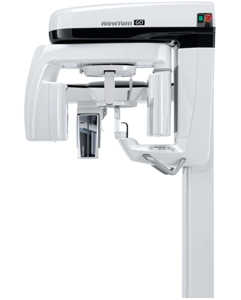
Go
2D/3D CEPH Integrated Imaging
-
 Broad diagnostic potential
 -
 Accessible technology
 -
 Maximum connectivity
 -
 Minimal radiation dose
IMAGING EXCELLENCE COMBINED WITH THE VERSATILITY OF A COMPLETE AND SAFE, TECHNOLOGICALLY ADVANCED SYSTEM.
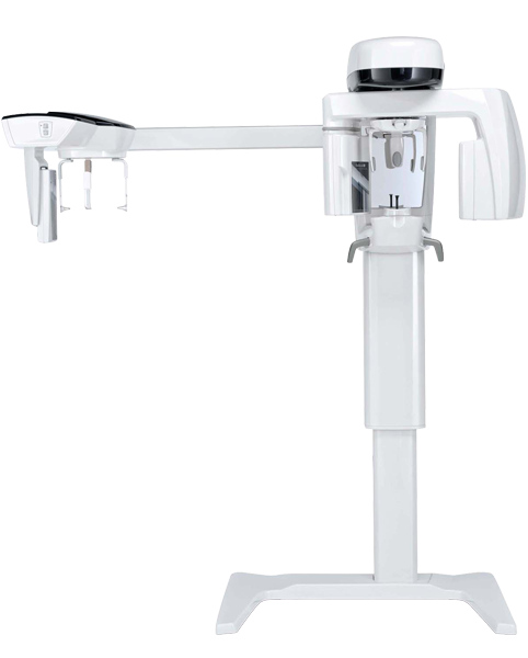
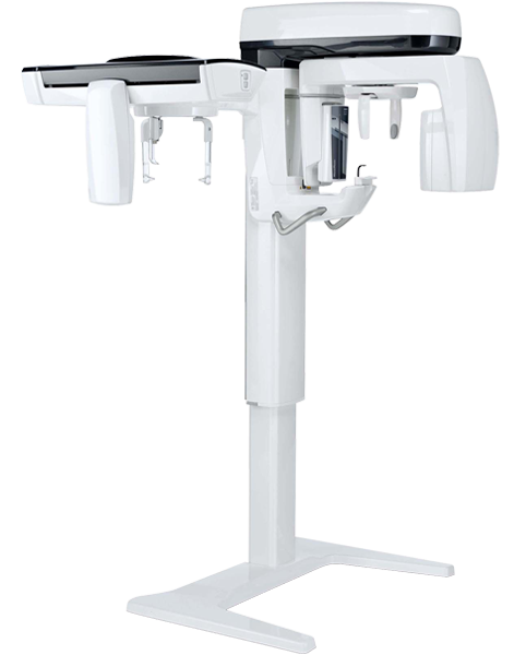
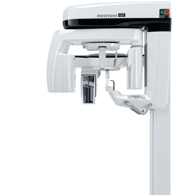
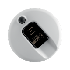
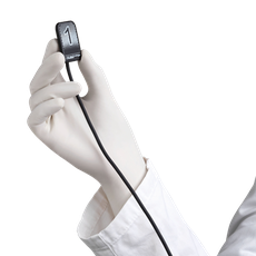
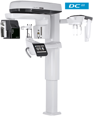
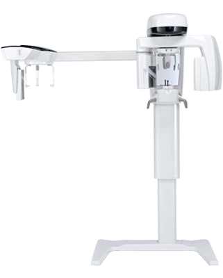
GO 2D/3D/CEPH is a flexible platform that comes ready for the optional integration of the teleradiographic arm in a 2D or 3D configuration. Able to provide high resolution images, the platform prioritises patient health thanks to low exposure protocols and exclusive SafeBeamTM technology, which lets users adapt the dose to their actual diagnostic needs and the size of the scanned anatomical area. Excellent ergonomics and an adaptive alignment system ensure correct positioning of the patient and perfect focusing for clear, detailed images. A virtual control panel guides the operator through each stage of the examination. NNT is the technologically advanced software platform to manage, process, consult and share diagnostic images.
Diagnostic images
- HEAD & NECK
Diagnostic images
 Back
Back
HEAD & NECK
- DENTITION
- 2D Panoramic
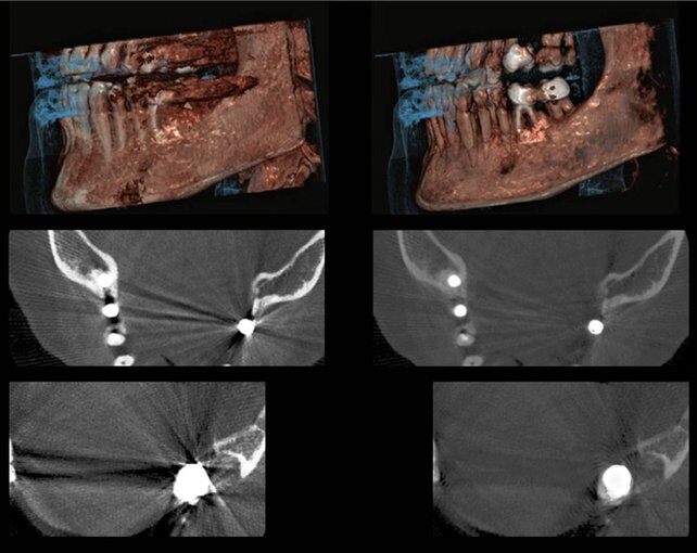
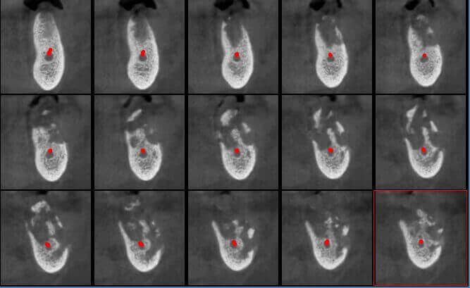
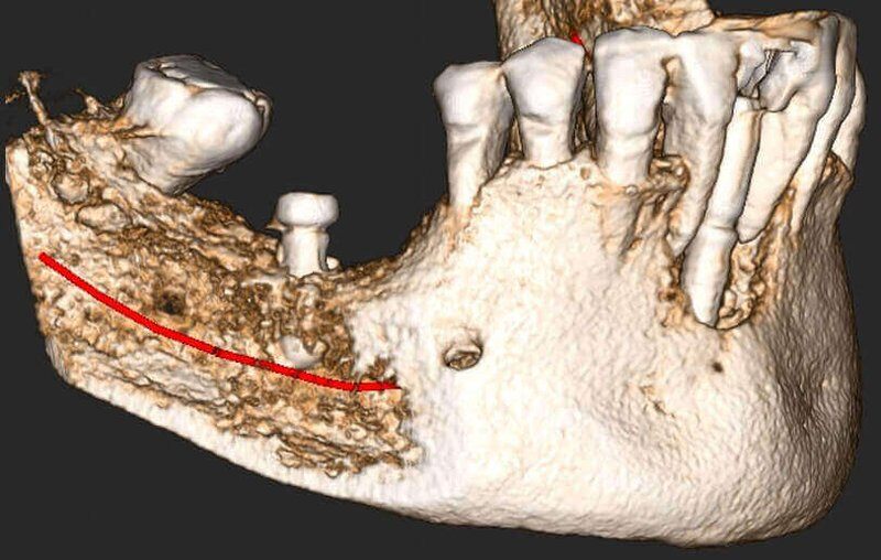
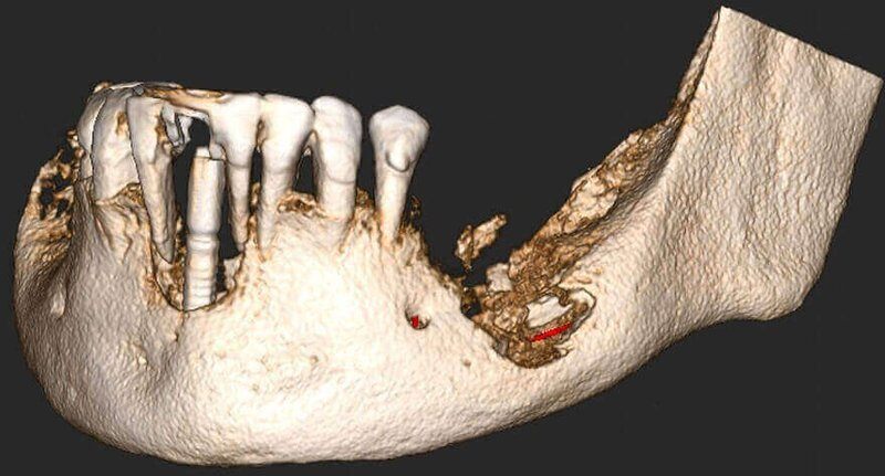
- Lower Jaw: Implants and Bone tumor. IAN evaluation
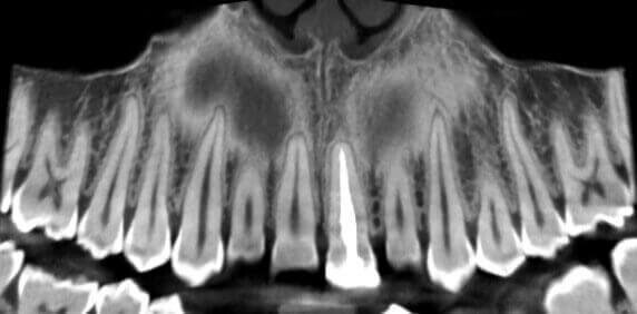
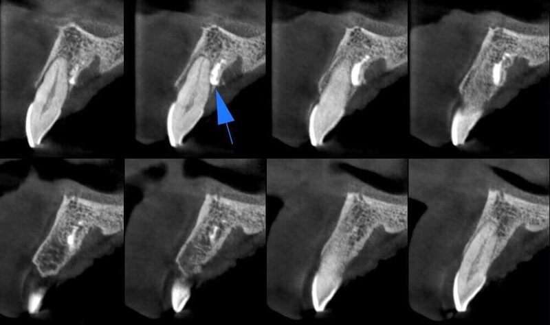
- Mesiodens
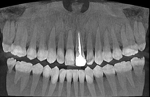
- Endodontic treatment in Incisor 2.1
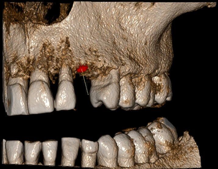
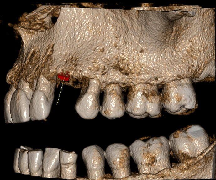
- Implant evaluation Q2
- TEMPOROMANDIBULAR JOINT
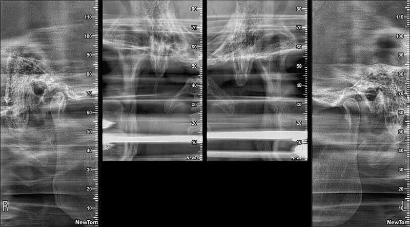
- TMJ Open / Closed mouth
- MAXILLARY SINUSES
- Maxillary Sinuses evaluation_2
2D Panoramic
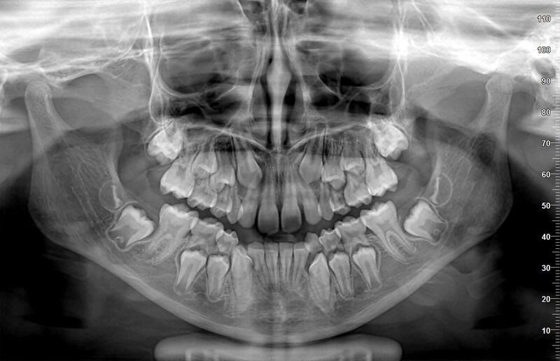
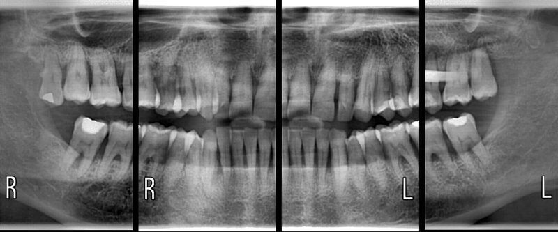
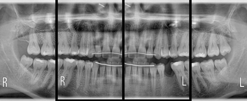
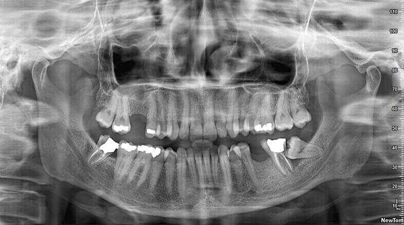
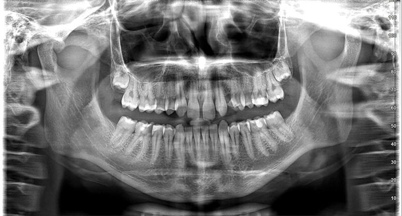
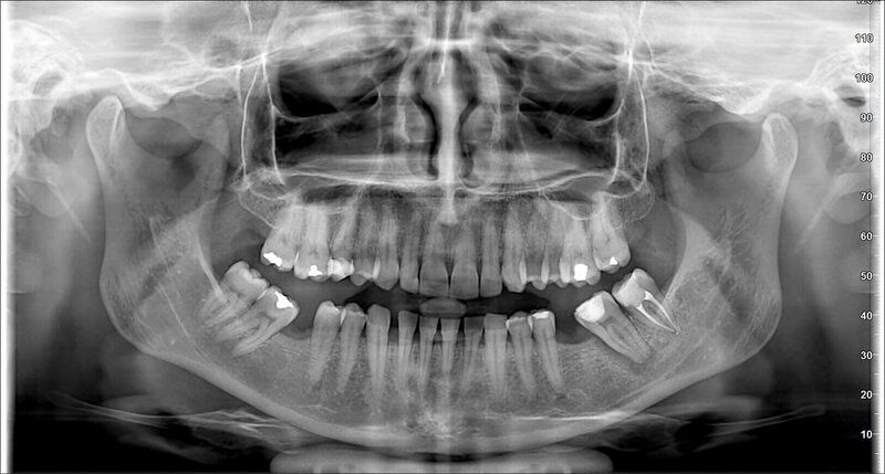
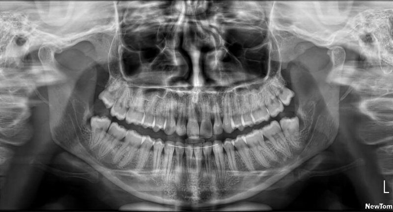
Lower Jaw: Implants and Bone tumor. IAN evaluation

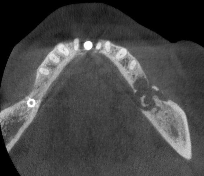
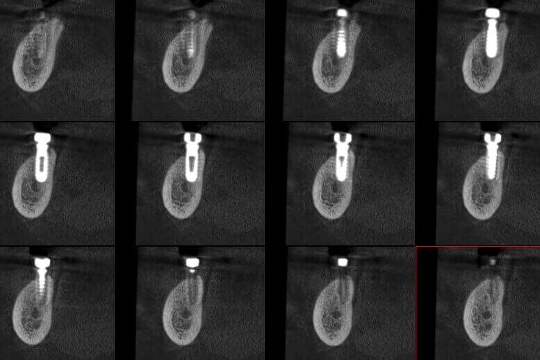
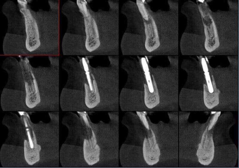
Mesiodens
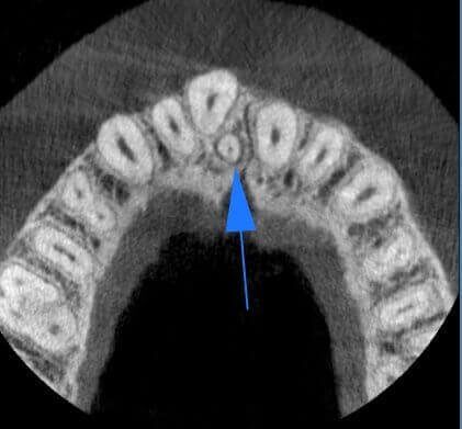
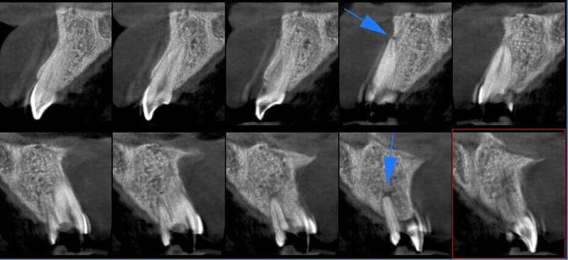
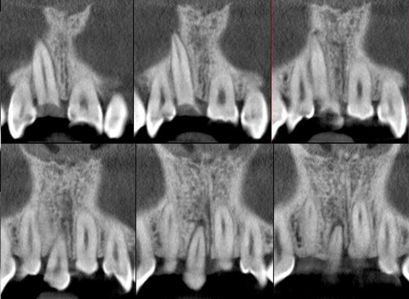
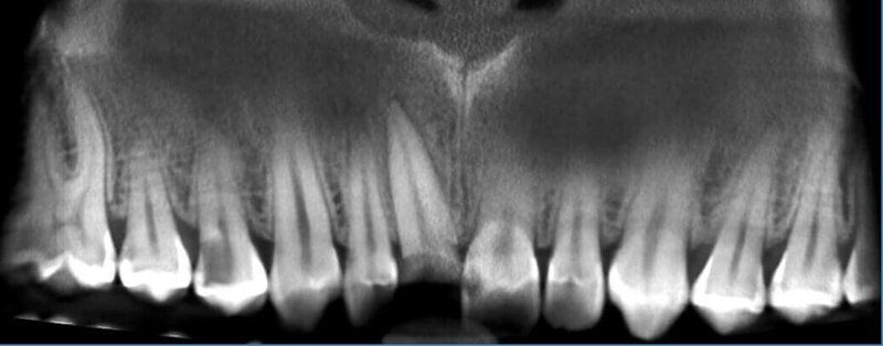
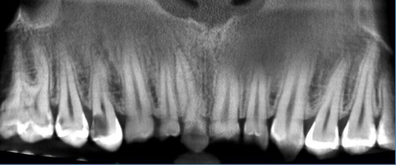
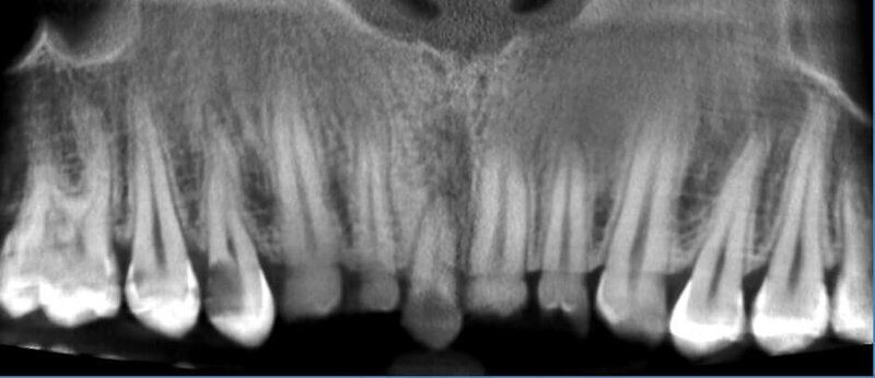
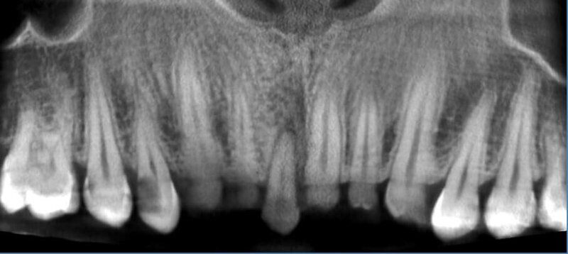
Endodontic treatment in Incisor 2.1
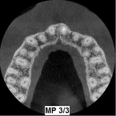
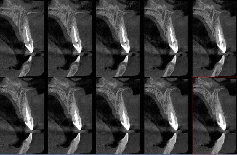
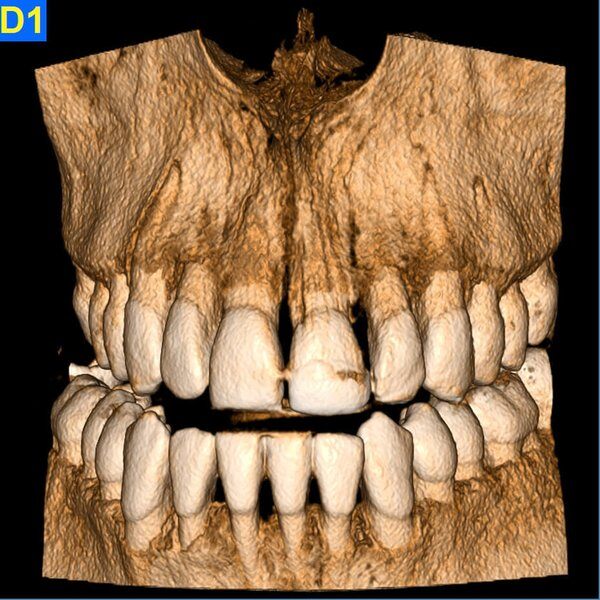
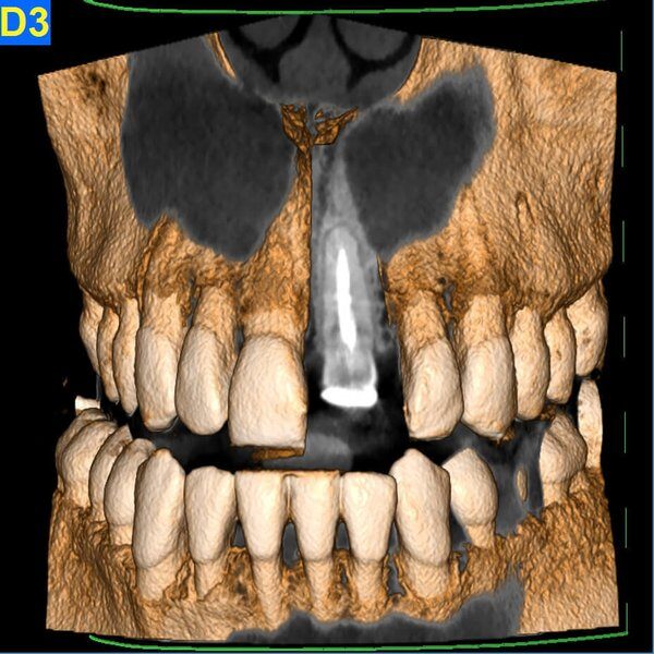
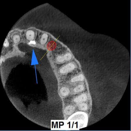
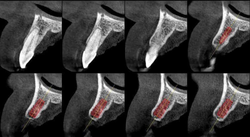
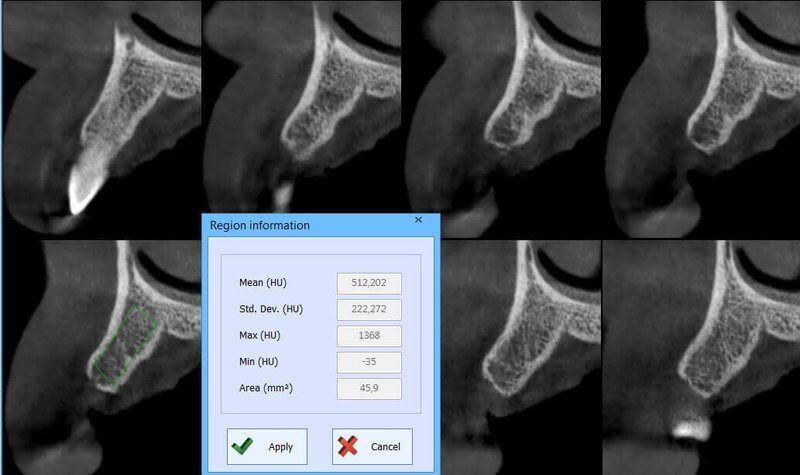
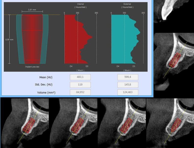
Implant evaluation Q2
TMJ Open / Closed mouth
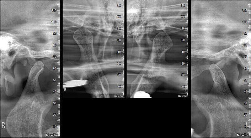
Maxillary Sinuses evaluation_2
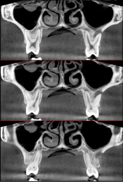
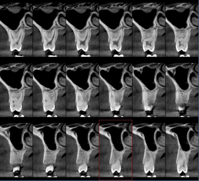
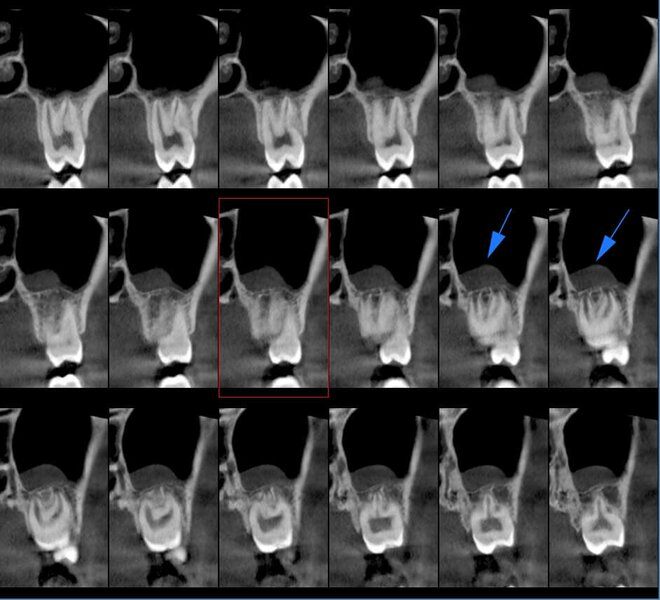
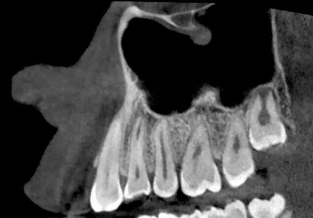
Solutions developed to maximise examination quality, from positioning systems to automated collimation.
GO 2D/3D has a single native 16-bit sensor that produces 2D and 3D images with thousands of grey levels. Image quality is ensured by advanced algorithms and protocols and by high- tech image sequencing. The high frequency, pulsed- emission generator adjusts exposure to obtain the best scans with a minimum dose.
Moreover, the cephalometric exam collimation system is based on automatic movement of the turret, which rotates and lowers the sensor, creating an opening for the X-rays directed at the 2D sensor on the teleradiographic arm.
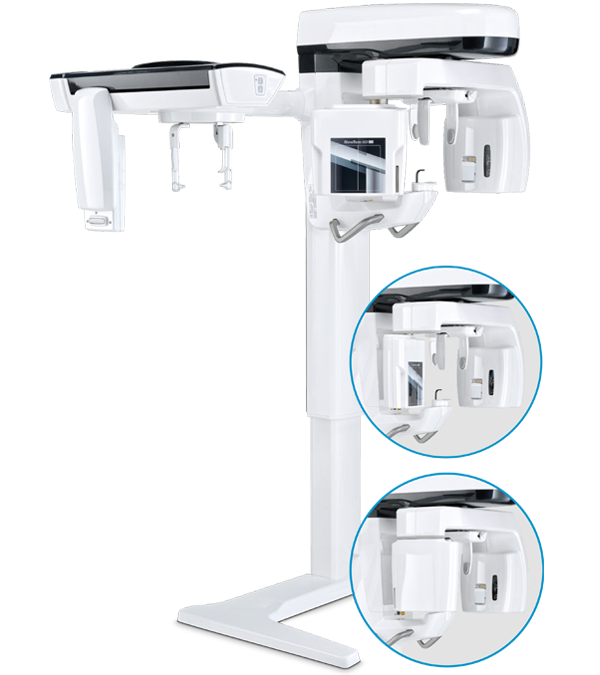
Top quality 2D imaging obtained through many advanced functions for more effective diagnostics.
It supplies detailed images thanks to the sensitivity of the newly developed CMOS sensor. GO 2D/3D offers quick and accurate diagnoses with several image acquisition software options designed to obtain high quality 2D images for all diagnostic needs. Excellent, clear and detailed panoramic images with ApT (Autoadaptive picture Treatments) technology. The aPAN (adaptive PAN) function allows five layers of panoramic images to be captured in a single scan in order to choose the most suitable one for the scope of the examination.
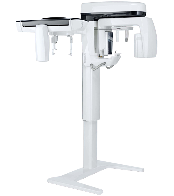
Clinical potential extended to meet all 2D diagnostic requirements through the CEPH arm.
Compact and available with relocatable PAN-CEPH sensor, the CEPH extension is equipped with a dedicated head support unit with two available side rod lengths. The CEPH application can be integrated at the time of purchase, but also retrofitted on equipment supplied in CEPH Ready version. All examinations are performed according to dedicated protocols for adults and children, optimised to reduce patient exposure based on actual scanning requirements. Accurate assessments before the application of dental braces, temporomandibular joint (TMJ) and maxillary sinus imaging, lateral and frontal teleradiographs.
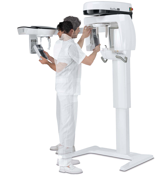
Detail-rich volumes for every clinical need while safeguarding patient health.
NewTom GO generates outstanding volumetric images and for each FOV, ranging from 6 x 6 to 10 x 10 cm. A choice of 3 protocols allows the required X-ray dose to be adapted to specific needs: from very low for quick scans required by surgical follow-up checks, through regular for treatment planning, to a very high level of detail for the analysis of micro-structures.
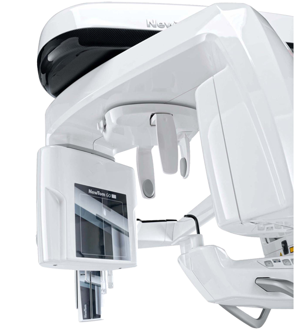
Advanced protocols and systems to reduce the radiated dose to a minimum.
Top quality imaging with a very low dose of radiations. Protocols defined by NewTom research in over 20 years of experience allow to automatically adapt exposure based on the anatomical characteristics of the patient, on the anatomical district examined and on actual diagnostic needs.
- ECO CEPH
- ECO SCAN AND ADAPTIVE FOV
- ECO PAN AND VARIABLE COLLIMATION
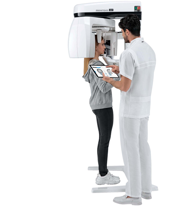
Excellent comfort for fast and stable positioning of the patient.
Designed to ensure excellent positioning of the patient, GO 2D/3D allows to rapidly find the correct position for examinations that are always perfect. The self-adaptive functions of GO 2D/3D allow to perform accurate examinations with top quality, diagnostically valuable images.
The operator has tools for patient positioning and guided alignment to obtain perfect focusing.
Features and specs
- Single sensor
- 2D High quality and Practical
- CEPH Arm
- Excellence in 3D
- Minimum dose, maximum diagnostic quality
- Ergonomics
Single sensor
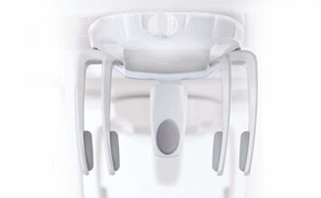
With its five contact points, the 3D scan head support helps staff position the patient correctly and comfortably.
Frontal and lateral contact points can be adjusted to maximise both patient stability during the scan and, consequently, the quality of the obtained data.
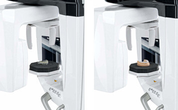
A specific protocol allows for tomographic scans of radiological templates, prostheses, models or impressions after they have been positioned on a special support.
2D High quality and Practical
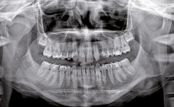
ORTHOGONAL PANORAMIC FUNCTION
The adaptive PAN function provides, in a single scan, 5 optimised images from which users can choose the panoramic view that best suits their diagnostic needs. Captured orthogonally, the dental arch image clearly highlights interproximal spaces and the entire root structure without any overlap
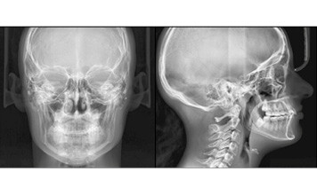
NEW CEPH HR FUNCTION
The highly compact teleradiographic arm completes the available 2D functions with a wide range of CEPH tests carried out with dedicated protocols for high-resolution imaging. With collimation designed to reduce X-ray doses and quick scan times the focus is on the patient’s health.
Image sequence:
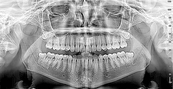
ADULT PANORAMIC IMAGING
Standard panoramic software provides a complete, accurate view of the dental arches, maxillary sinuses and temporomandibular joints. The integrated feature of panoramic view orthogonal capturing perfectly highlights interproximal spaces and the entire root structure without any overlap.
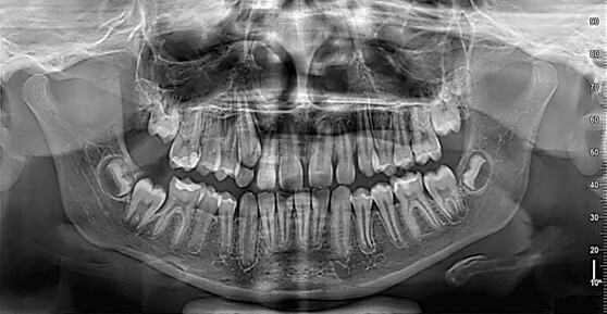
CHILD PANORAMIC IMAGING
Child panoramic imaging with vertical collimation and low radiated dose: field of view and exposure are adapted to the paediatric patient’s build.
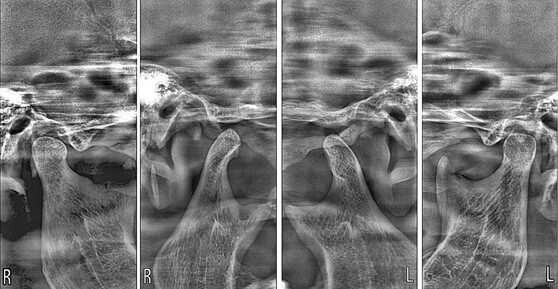
TEMPOROMANDIBULAR JOINT
The trajectories dedicated to the temporomandibular joints (TMJ) generate four projections with a single scan: two lateral and two postero-anterior, with mouth either open or closed.
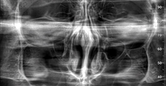
MAXILLARY SINUSES
The SIN software uses a focal layer that has been specially designed to improve maxillary sinus examinations. A dedicated support allows to obtain both frontal and lateral slices.
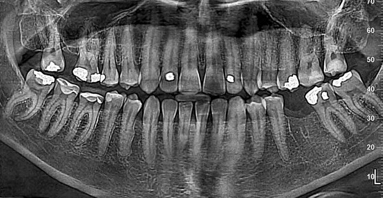
DENTITION
Clear detailed images limited only to the teeth, either whole or partial, with orthogonal projection and better signal-noise ratio. Ideal for periodontal controls.
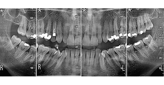
BITEWING
Optimised collimated interproximal projection with a low dose to study dental crowns. An alternative to intraoral bitewings, with a less invasive and more comfortable procedure.
CEPH Arm
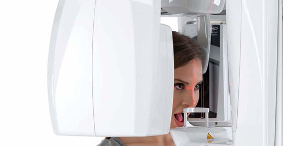
HEAD SUPPORT UNIT
The head support unit, which includes four partly adjustable contact points, guides the patient into the correct position for every kind of examination, including TMJ and maxillary sinus scanning.
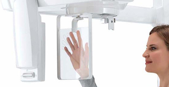
CARPAL
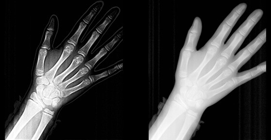
The teleradiographic module includes a convenient support for carpal scanning.
Image sequence:
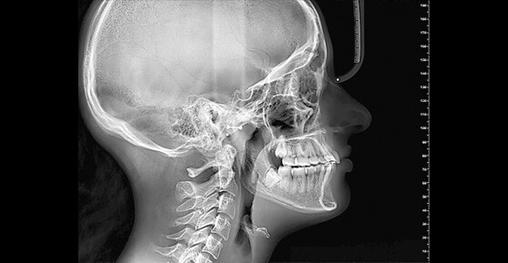
LATERAL CRANIAL TELERADIOGRAPHY
Through lateral projections, detailed examinations of the bone structures with highlighted soft tissues are obtained, critically important for cephalometric studies. Try the innovative CEPH-X online service for automatic cephalometric tracing based on an artificial intelligence algorith.
FRONTAL CRANIAL TELERADIOGRAPHY
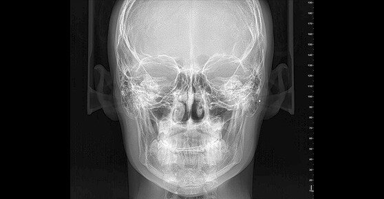
For the purpose of completing each treatment correctly, frontal projections can be used to scan for asymmetries and malocclusions.
CARPAL TELERADIOGRAPHY
Residual growth potential assessment through carpal examination. The dedicated support facilitates the correct performance of the scan.
ApT (AUTOADAPTIVE PICTURE TREATEMENTS)
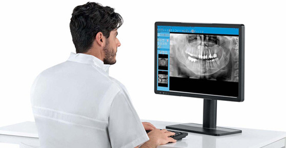
Auto-adaptive filters automatically improve every 2D image to ensure the best result for every projection.
Excellence in 3D

Complete child/adult superior arch
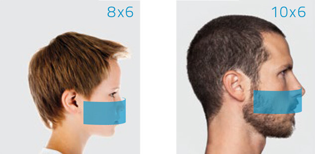
Volumes with FOV 8 x 6 cm and 10 x 6 cm produce images of localised anatomical districts, such as, for example, a maxillary sinus with suitable lift to insert an implant. The ideal solution in the field of implantology to assess both implant site and bone density.

Complete child/adult lower arch
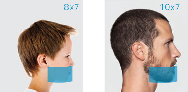
The 8 x 7 cm and 10 x 7 cm FOVs are designed for analyses of the mandibular region. In the case of impacted canines, where it is necessary to assess their relationship with the mandibular canal and adjacent anatomical structures, the advanced image acquisition and processing functions allow to easily and rapidly highlight the slices of interest.

Upper and lower local investigations
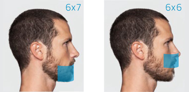
With FOVs 6 x 7 cm and 6 x 6 cm, scans can be performed with very high resolution to clearly see even the smallest detail. This mode is especially indicated for endodontic and periodontic applications.

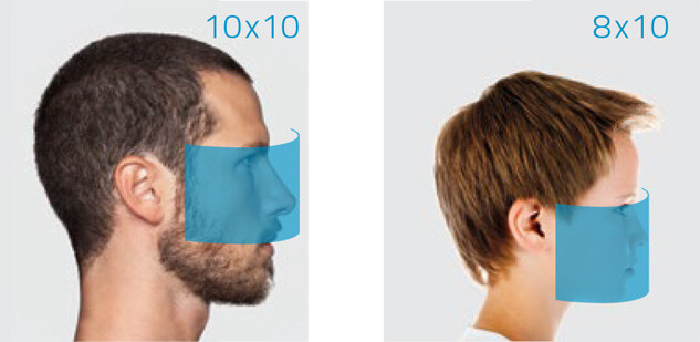
Studying adult/child maxillary sinuses

Minimum dose, maximum diagnostic quality
Combining technology, performance and safety
ECO CEPH
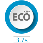
Given the nature of a cephalometric examination, often used in cases of pedodontics, NewTom has developed a protocol that minimises the X-ray dose to which the patient is exposed. With a scan time limited to just 3.7 seconds, the patient benefits from minimal X-ray exposure and an extremely short time inside the device. In addition to the scanning mode, the longer ear guards protect the child’s thyroid from unnecessary exposure during the examination.
ECO SCAN AND ADAPTIVE FOV
NewTom, ever keen on patient health, was the first to use pulsed emission with CBCT technology applied to dental imaging, thus considerably reducing the dose of radiations emitted during 3D examinations. The introduction of the 3D ECO SCAN protocol (ultra rapid scan of only 6.4 seconds and actual emission time of only 1.6 seconds) provides the ideal solution for post-surgery follow-up examinations and for all situations where the X-ray dose must be kept to a minimum. Instead, the 3D aFOV (adaptive FOV) function allows the size of the radiated anatomical district to be limited in order to adapt to the different morphological features of adults and children or to simply perform sectional examinations up to a 6 x 6 cm FOV, with a minimum effective dose in ECO mode of 9 μSv.
ECO PAN AND VARIABLE COLLIMATION
GO 2D/3D offers several PAN software options with variable collimation for adults and children, dedicated image acquisition for the dentition area only and bitewing views.
The ECO PAN protocol allows an ultra rapid scan to be performed (6.6 seconds) to further reduce the radiation dose down to 5 μSv. Versatile, high quality 2D diagnoses with limited emission.
Ergonomics
GUIDED ALIGNMENT
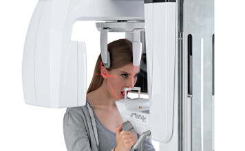
Three laser guides and a wide front mirror allow quick and precise positioning of the patient. The device can be controlled by the operator via a user-friendly on-board keyboard or by using the dedicated App.
ALIGNMENT CHECKS
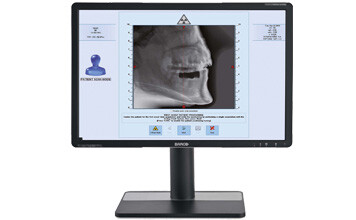
Before performing a 3D scan, two scout images allow to precisely check and adjust patient alignment via PC-controlled servo-assisted movements.
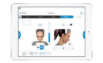
VIRTUAL CONSOLE
Rapid and user-friendly image acquisition with the virtual console on PC or a dedicated software for iPad. The operator follows all examination phases, from the choice of an examination to scan start.

NNT. ADVANCED SOFTWARE FUNCTIONS.
Extensive sharing and processing power with the ultimate imaging platform.
NewTom’s NNT software offers all functions required to perform, process, display and share 2D and 3D examinations. NNT also provides different application modes and functions specifically intended to plan the best treatment for implantology, endodontics, periodontics and radiology applications as well as maxillofacial surgery.



 Download Brochure
 video
video
 Find a dealer
 Contact us
