
Knee joint

Wrist rotation
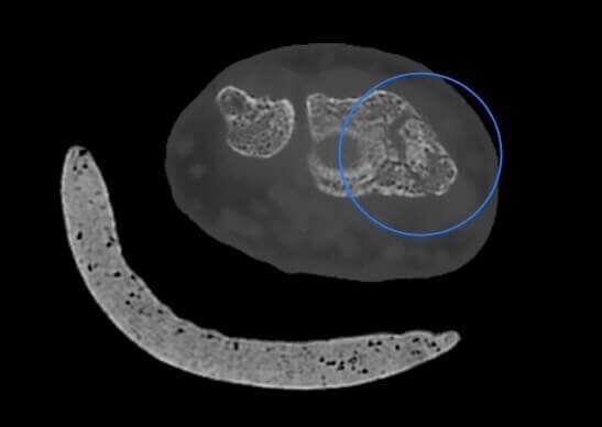
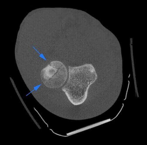
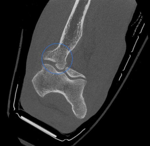
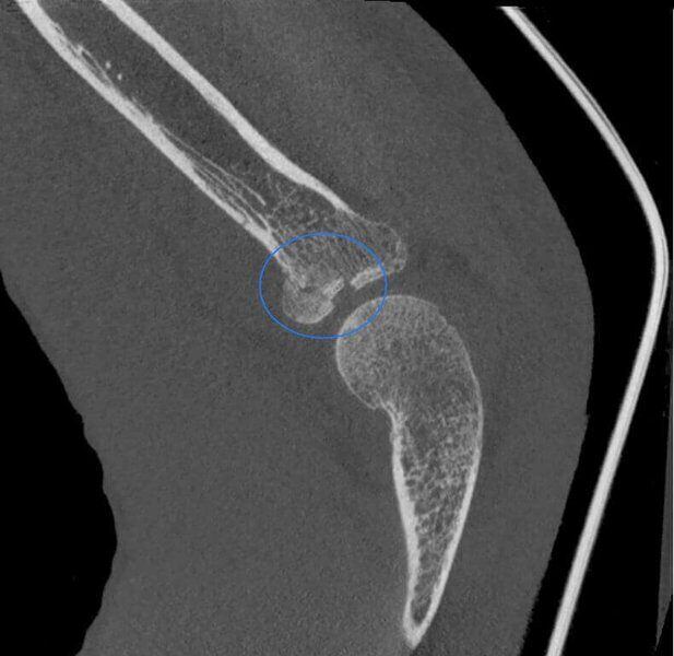
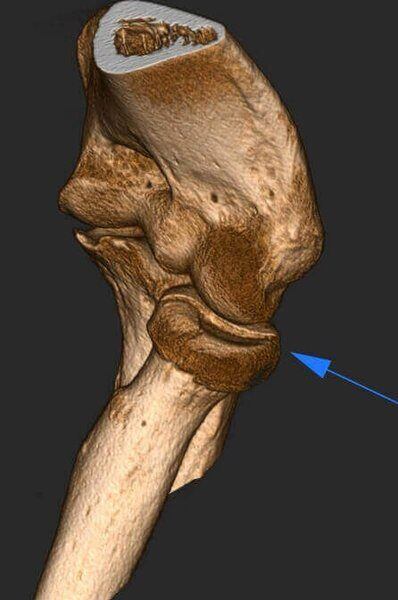
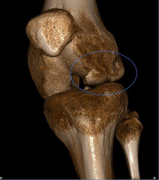
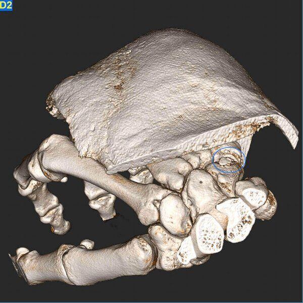
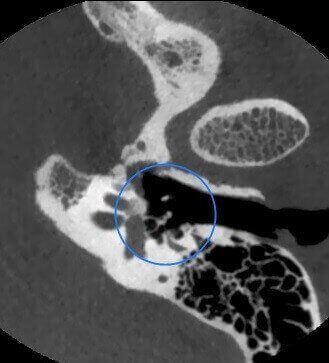
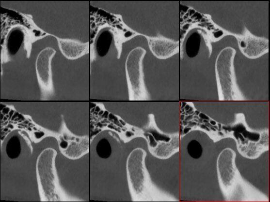
Coronoid process of Ulna fracture

Fibula Distal Fracture
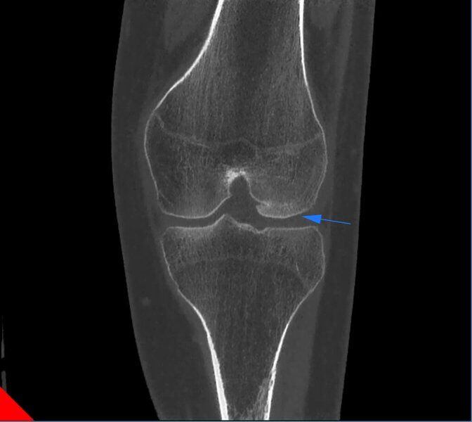
Osteocondral lesion in patient with ostesynthesis tool
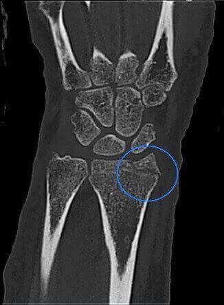
Radius Styloid process fracture
Radius Styloid process fracture
Radius Styloid process fracture
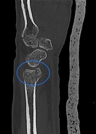
Radial Head fracture in patient with cast
Radial Head fracture in patient with cast
Radial Head fracture in patient with cast
Radial Head fracture in patient with cast
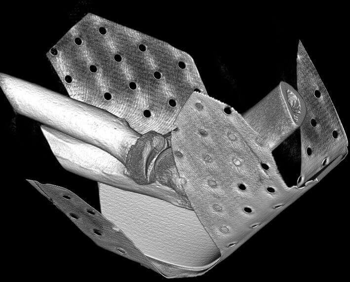
Radial Head fracture in patient with cast
Radial Head fracture in patient with cast
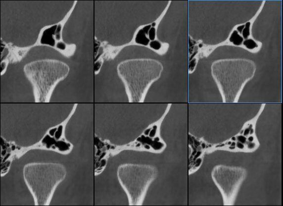
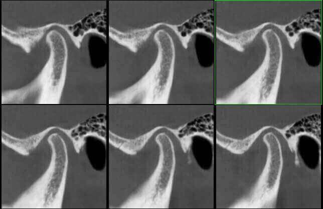
Scaphoid fracture

Scaphoid fracture

Scaphoid fracture
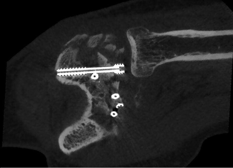
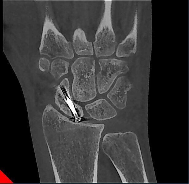
Scaphoid fracture with Ostesynthesis tool
Scaphoid fracture with Ostesynthesis tool
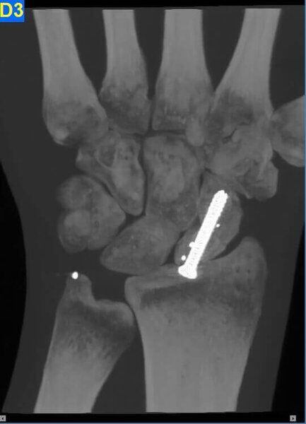
Scaphoid fracture with Ostesynthesis tool

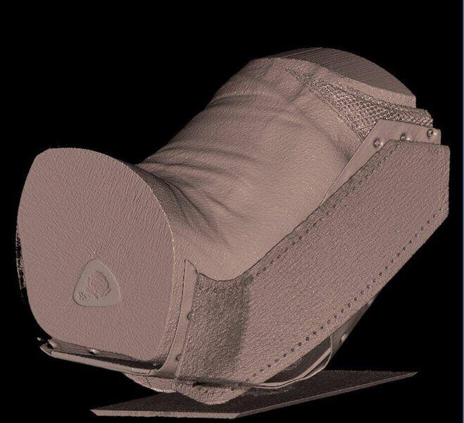
Wrist with Plaster Cast

Wrist with Plaster Cast
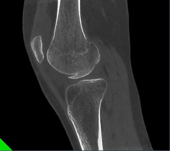
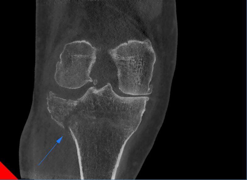
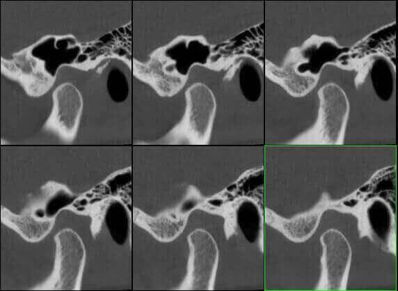
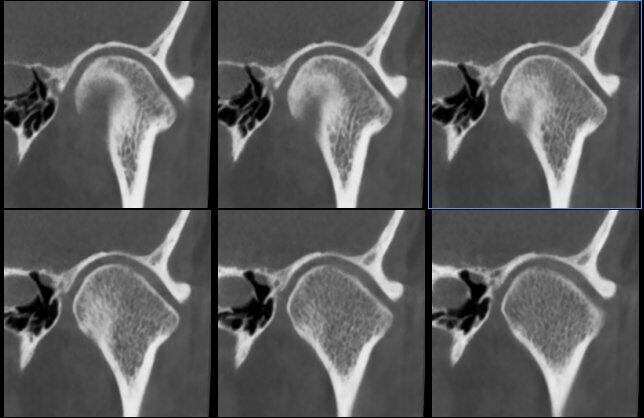
Lateral femoral condile fracture
Lateral femoral condile fracture

Lateral femoral condile fracture
Lateral femoral condile fracture
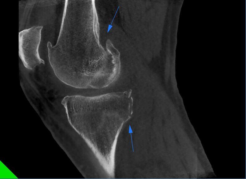
Knee Multiple fractures
Knee Multiple fractures
Knee Multiple fractures

Knee

Knee

Bone growth

Bone growth

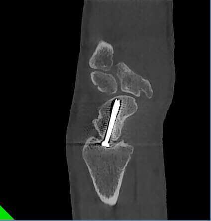
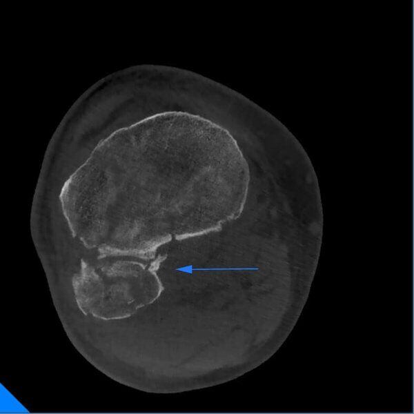
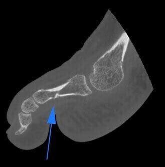
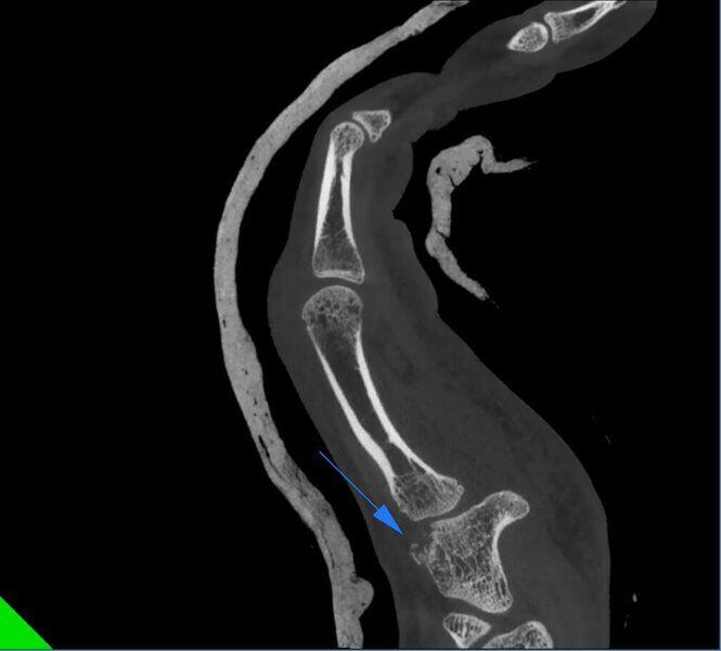
Sesamoid fracture

Sesamoid fracture
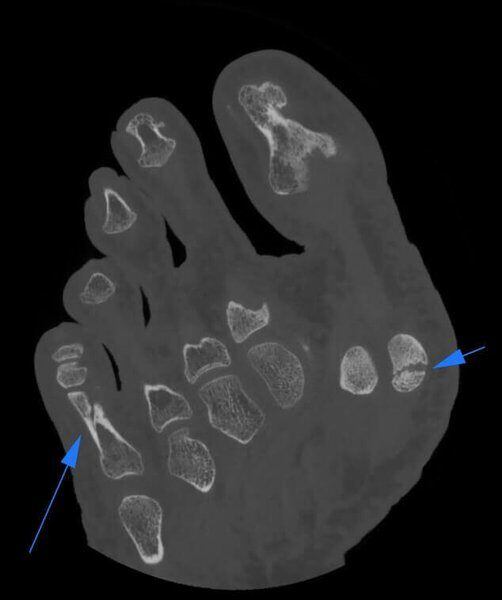
Sesamoid and Phalange fracture
Sesamoid and Phalange fracture
Sesamoid and Phalange fracture
Sesamoid and Phalange fracture
Sesamoid and Phalange fracture

Scaphoid microfractures

Scaphoid microfractures

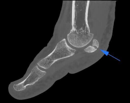
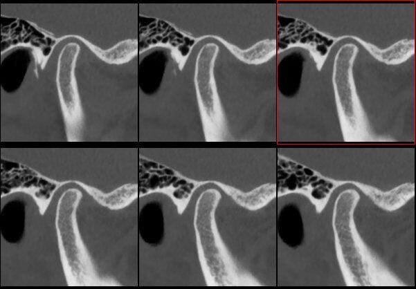
Os Trigonum

Os Trigonum

Os Trigonum

Heel Osteonecrosis

Heel Osteonecrosis

Heel Osteonecrosis

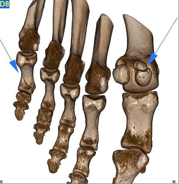
Missing fingers

Missing fingers
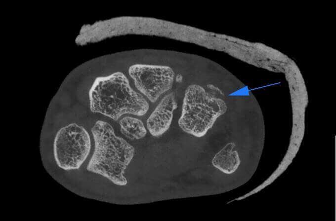
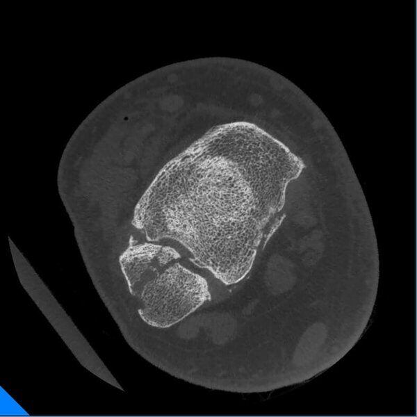
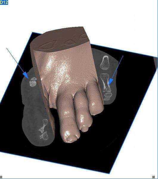
Unciform bone microftracture
Unciform bone microftracture
Unciform bone microftracture

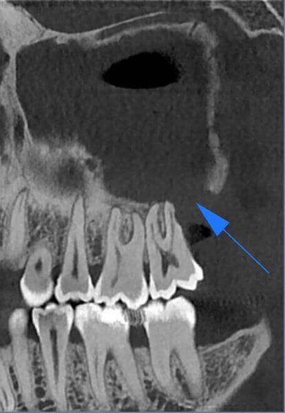
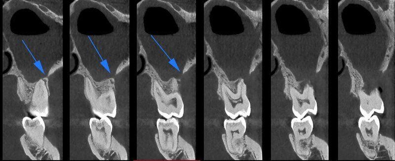
Cisti mandibolare in Q4

Cisti mandibolare in Q4

Cisti mandibolare in Q4

Cisti mandibolare in Q4

Cisti mandibolare in Q4

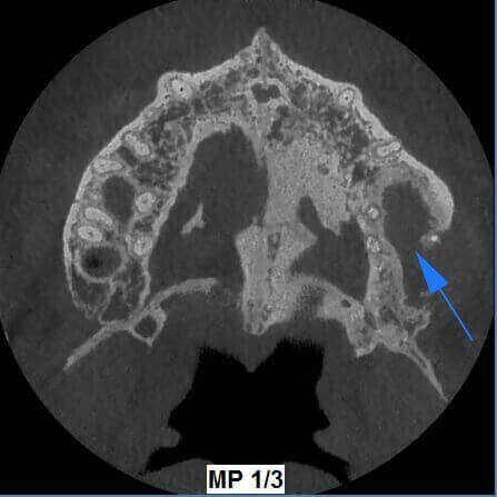
Doppia Arcata

Doppia Arcata

Doppia Arcata

Doppia Arcata

Doppia Arcata

Doppia Arcata

Deglutizione
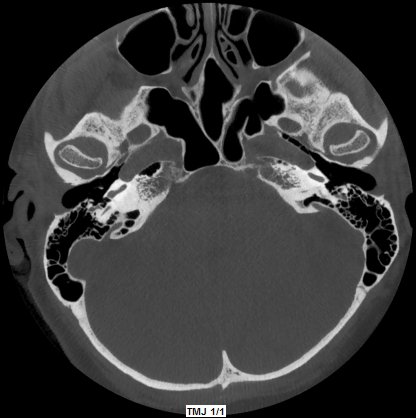
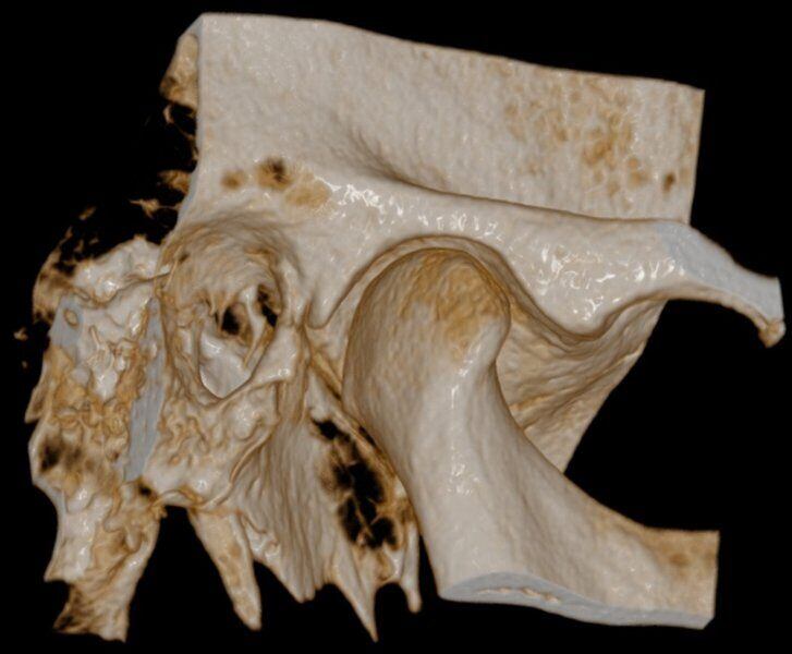
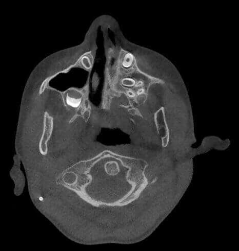
Catena Ossiculare e Nervo Facciale
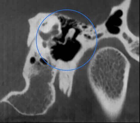
Catena Ossiculare e Nervo Facciale
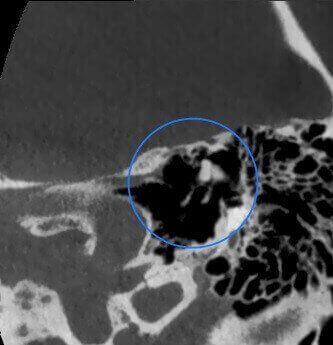
Catena Ossiculare e Nervo Facciale
Catena Ossiculare e Nervo Facciale

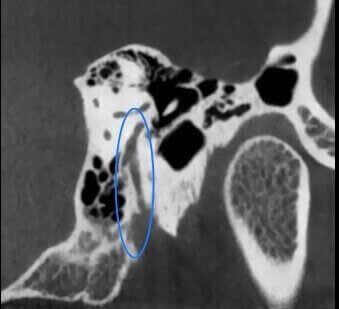
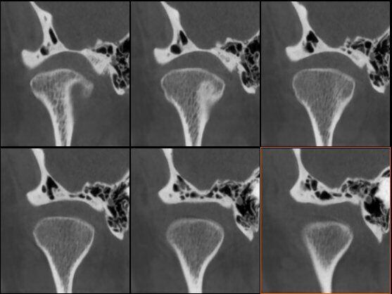
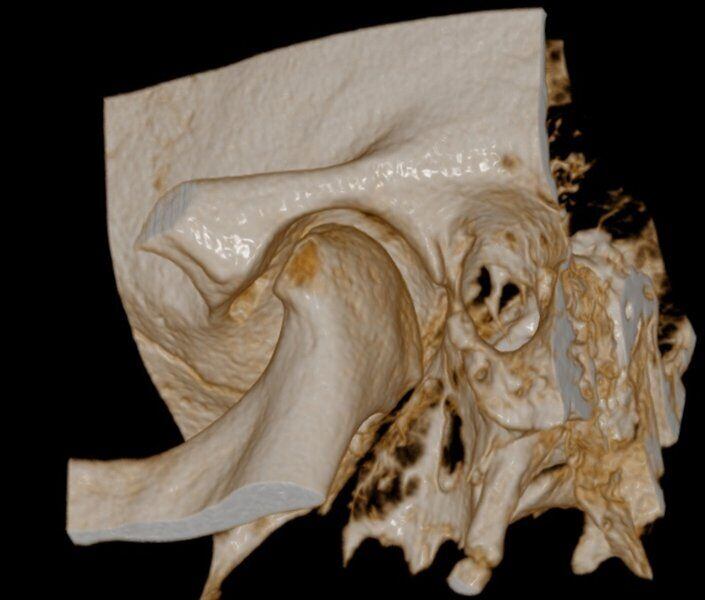
Impianto Cocleare

Impianto Cocleare

Impianto Cocleare

ATM Bocca Aperta
ATM Bocca Aperta
ATM Bocca Aperta
ATM Bocca Aperta
ATM Bocca Aperta
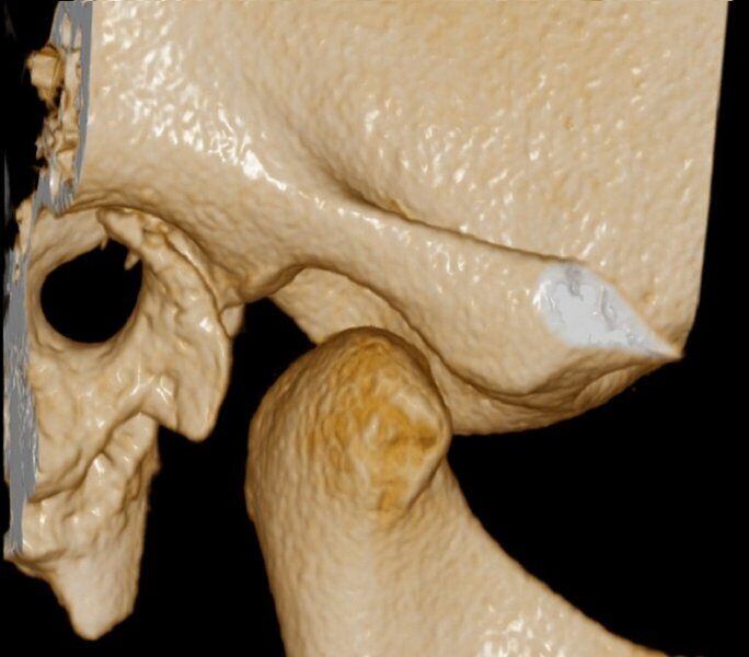
ATM Bocca Aperta
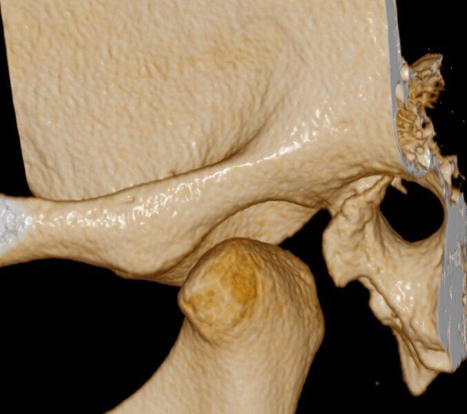
ATM Bocca Aperta
ATM Bocca Chiusa
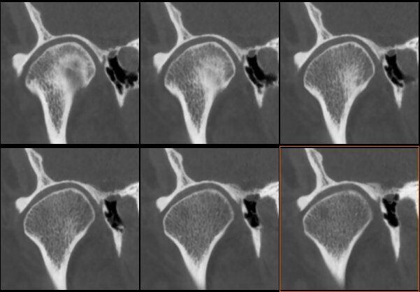
ATM Bocca Chiusa
ATM Bocca Chiusa
ATM Bocca Chiusa
ATM Bocca Chiusa
ATM Bocca Chiusa
ATM Bocca Chiusa
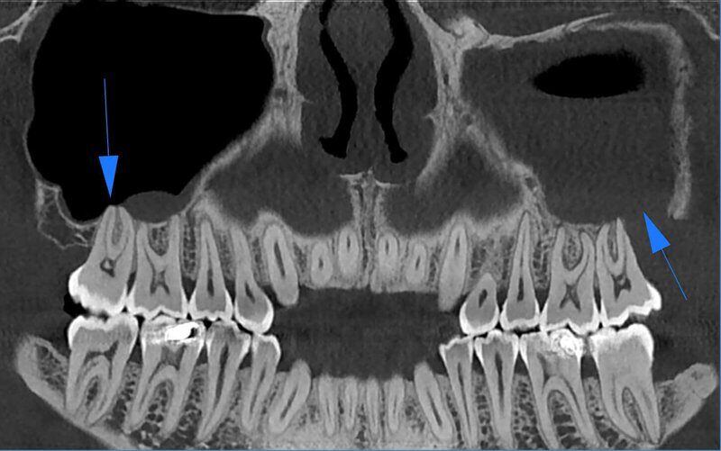
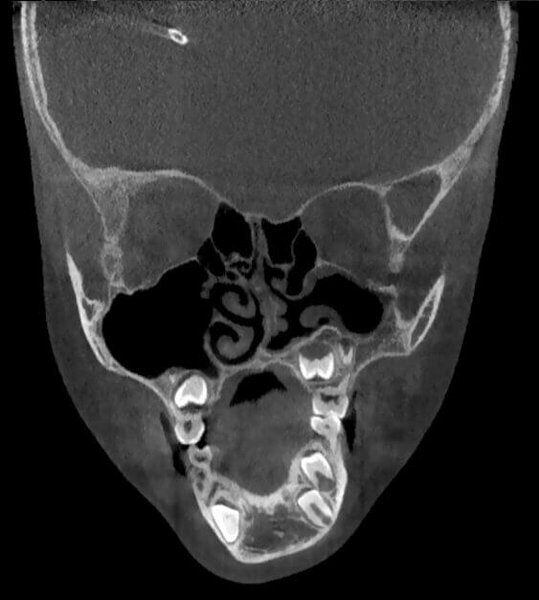
Rottura del pavimento del Seno Mascellare sinistro con Sinusite
Rottura del pavimento del Seno Mascellare sinistro con Sinusite
Rottura del pavimento del Seno Mascellare sinistro con Sinusite
Rottura del pavimento del Seno Mascellare sinistro con Sinusite
Acquisizione Maxiillofacciale per valutazione chirurgica di paziente con Sindrome di Goldenhar
Acquisizione Maxiillofacciale per valutazione chirurgica di paziente con Sindrome di Goldenhar
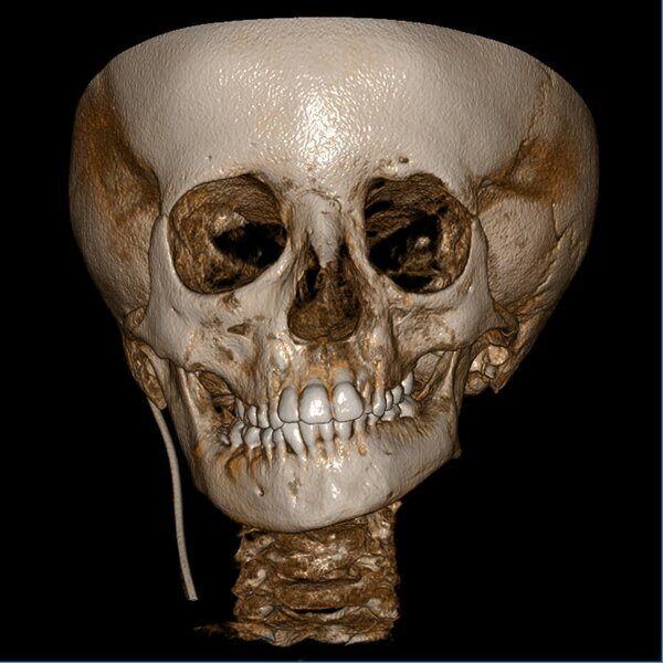
Acquisizione Maxiillofacciale per valutazione chirurgica di paziente con Sindrome di Goldenhar
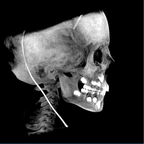
Acquisizione Maxiillofacciale per valutazione chirurgica di paziente con Sindrome di Goldenhar
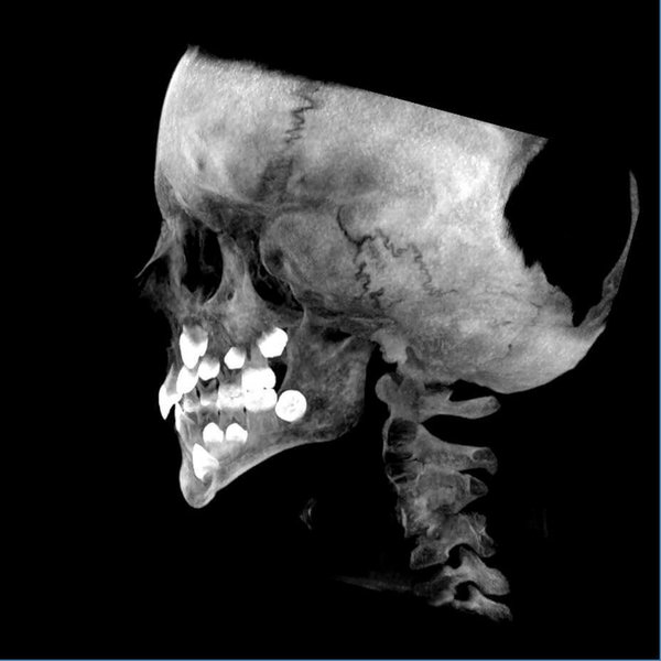
Acquisizione Maxiillofacciale per valutazione chirurgica di paziente con Sindrome di Goldenhar
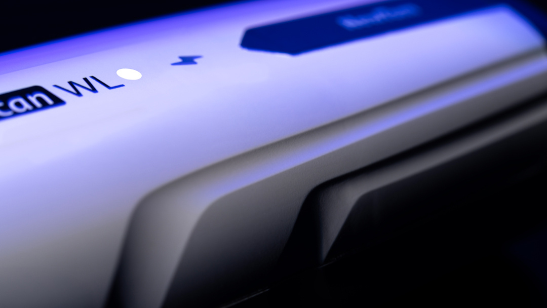
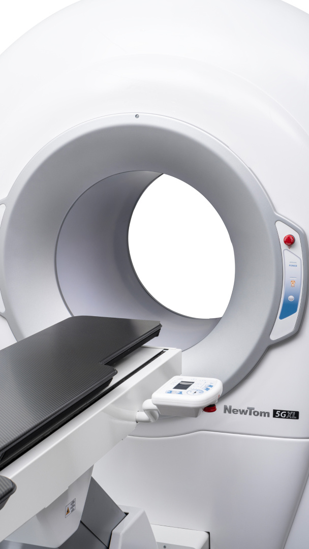
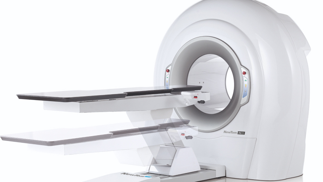
3D scans: precision, safety and maximum efficiency
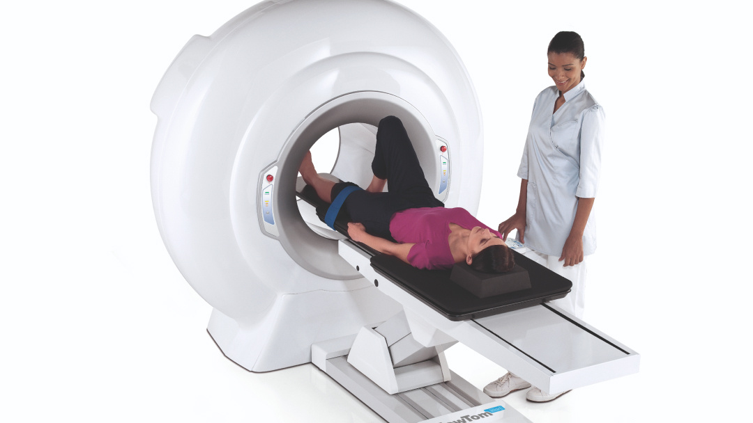
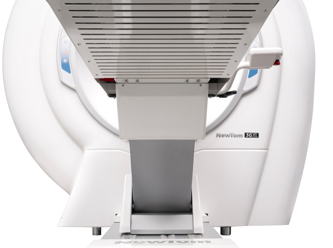
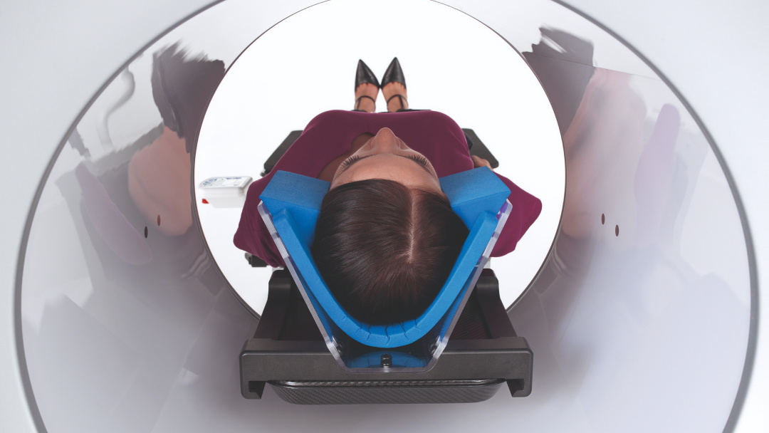
NewTom 5G XL takes 3D imaging to a new level, delivering ultra-high-resolution volumetric images with minimal radiation exposure. With the patient in a lying-down position, this device ensures optimal stability, reducing the risk of movement-induced artifacts and improving diagnostic quality.
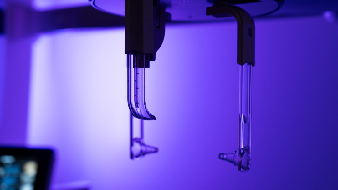
Advanced CBCT technology enables detailed bone tissue imaging with native isotopic voxel, non-overlapping slices and fewer artifacts, making of 5G XL the ideal solution for applications in dentistry, maxillofacial surgery, orthopedics, and ENT.
The wide FOV range, from 6x6 cm up to 21x19 cm, ensures maximum flexibility to meet every diagnostic need, while 360° scanning captures the entire volume in a single rotation, optimising scan times.
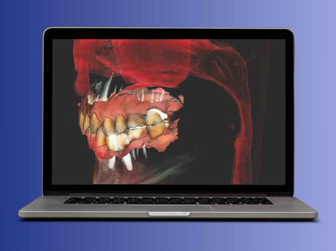
To aid image management, NNT - Medical Suite software has intuitive tools to capture, process and share 2D and 3D exams, with advanced features for treatment planning. Integration with CineX also allows moving anatomical structures to be analysed, while sophisticated reconstruction algorithms reduce artifacts and ensure sharp, detailed imaging in any clinical situation.
Focus on patient health
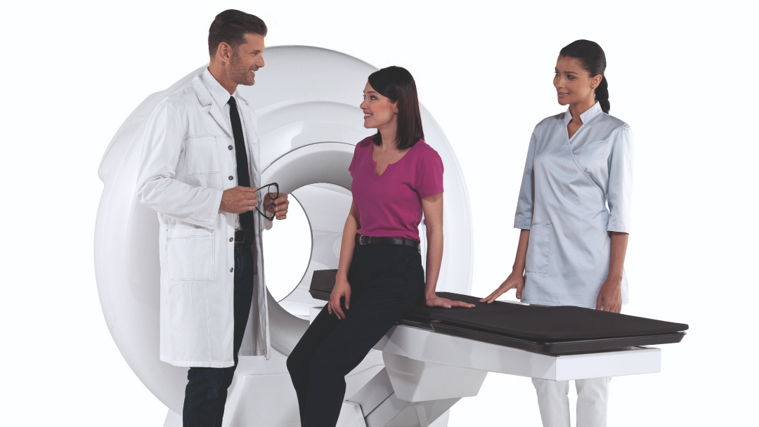
Wellbeing and safety are central to NewTom research.
5G XL offers top quality clinical examinations by delivering the lowest possible X-ray dose to patients based on diagnostic needs.
This is possible thanks to a number of technological solutions adopted.
5G XL offers top quality clinical examinations by delivering the lowest possible X-ray dose to patients based on diagnostic needs.
This is possible thanks to a number of technological solutions adopted.