5g-xl-vet-header

Characteristics

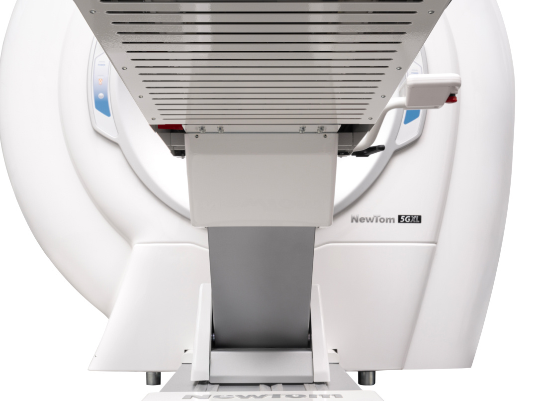
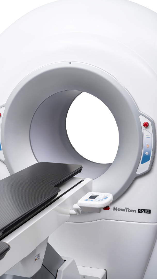
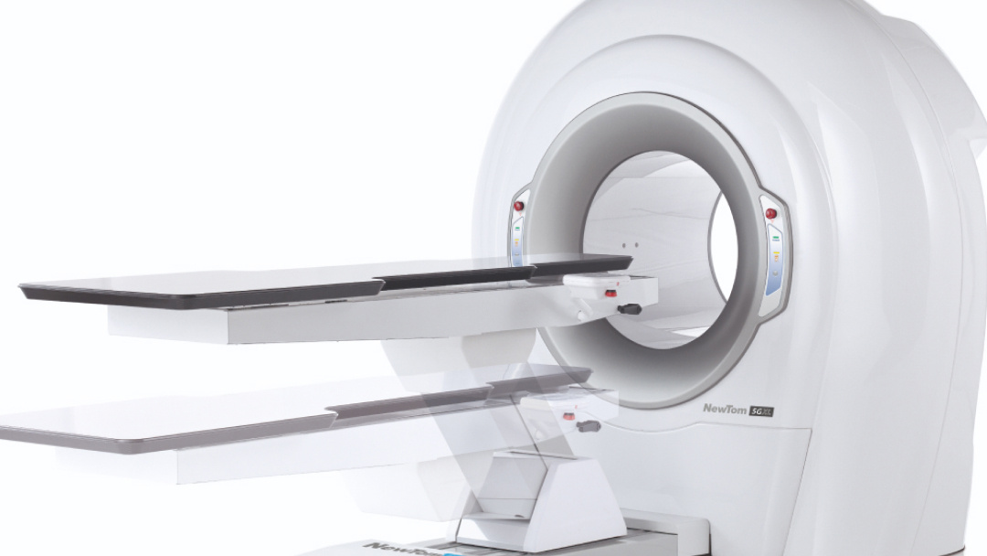
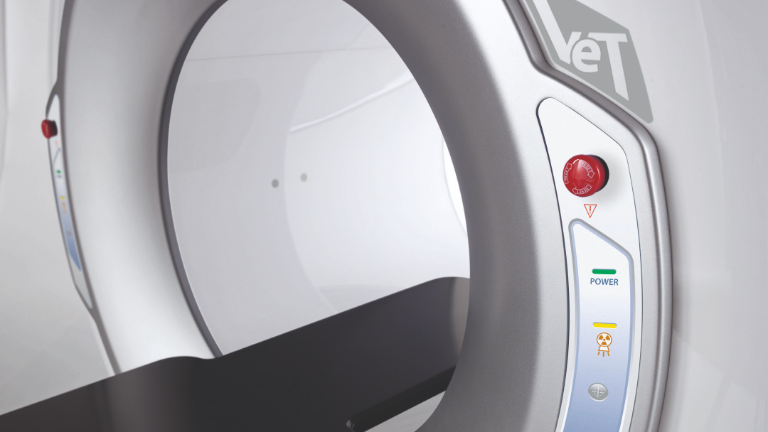
NewTom 5G XL VET is the CBCT device featuring a signature patient lying-down positioning, designed to ensure maximum stability, superior diagnostic quality and optimal comfort.  

  • Ultra high resolution images

    Thanks to the advanced technology applied, extra high resolution 2D and 3D images are obtained with reduced radiation exposure (up to 10 times lower than in traditional CT). 
  • Superior analysis performance

    The wide FOV range up to 21 x 19 cm enables detailed analyses for applications in dentistry, orthopedics, ENT and maxillofacial surgery. SafeBeam™ technology automatically adapts the dose, while the ECO Scan mode optimises irradiation, ensuring patient safety. 
  • Maximum focus on the patient

    The open gantry and motor-driven patient table of 5G XL VET ensure easy access even to sedated or trauma patients.  

Diagnostic imaging

With 5G XL, NewTom uses CBCT technology for new veterinary applications. Very high quality 2D and 3D images with a broad range of FOVs and dedicated software. 

  • Diagnostic images
5G XL VET_Immagini Diagnostiche_Lussazione ATM_1

5G XL VET_Immagini Diagnostiche_Lussazione ATM_2

5G XL VET_Immagini Diagnostiche_Lussazione ATM_3

5G XL VET_Immagini Diagnostiche_Lussazione ATM_4

5G XL VET_Immagini Diagnostiche_Lussazione ATM_5

5G XL VET_Immagini Diagnostiche_Lussazione ATM_6

5G XL VET_Immagini Diagnostiche_Ernia del disco lombare_1

5G XL VET_Immagini Diagnostiche_Ernia del disco lombare_2

5G XL VET_Immagini Diagnostiche_Ernia del disco lombare_3

5G XL VET_Immagini Diagnostiche_Ernia del disco lombare_4

5G XL VET_Immagini Diagnostiche_Tumore nasale_1

5G XL VET_Immagini Diagnostiche_Tumore nasale_2

5G XL VET_Immagini Diagnostiche_Tumore nasale_3

5G XL VET_Immagini Diagnostiche_Tumore nasale_4

5G XL VET_Immagini Diagnostiche_Tumore nasale_5

5G XL VET_Immagini Diagnostiche_Tumore nasale_6

5G XL VET_Immagini Diagnostiche_Tumore nasale_7

5G XL VET_Immagini Diagnostiche_Lussazione della rotula pre e post-operatoria_1

5G XL VET_Immagini Diagnostiche_Lussazione della rotula pre e post-operatoria_2

5G XL VET_Immagini Diagnostiche_Lussazione della rotula pre e post-operatoria_3

5G XL VET_Immagini Diagnostiche_Lussazione della rotula pre e post-operatoria_4

5G XL VET_Immagini Diagnostiche_Lussazione della rotula pre e post-operatoria_5

5G XL VET_Immagini Diagnostiche_Lussazione della rotula pre e post-operatoria_6

5G XL VET_Immagini Diagnostiche_Lussazione della rotula pre e post-operatoria_7

3D scans: precision, safety and maximum efficiency

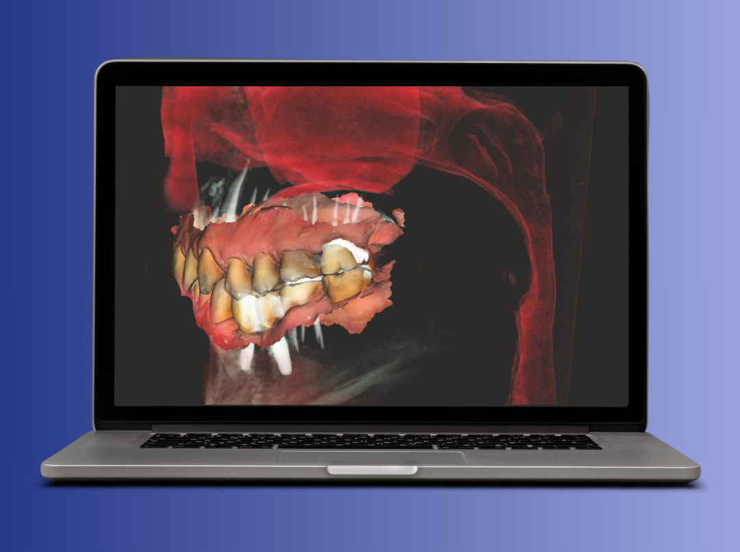
NewTom 5G XL VET takes 3D imaging to a new level, delivering ultra-high-resolution volumetric images with minimal radiation exposure. With the patient in a lying-down position, this device ensures optimal stability, reducing the risk of movement-induced artifacts and improving diagnostic quality. 

Advanced CBCT technology enables detailed bone tissue imaging with native isotopic voxel, non-overlapping slices and fewer artifacts, making of 5G XL the ideal solution for applications in dentistry, maxillofacial surgery, orthopedics, and ENT. 

The wide FOV range, from 6x6 cm up to 21x19 cm, ensures maximum flexibility to meet every diagnostic need, while 360° scanning captures the entire volume in a single rotation, optimising scan times. 

To aid image management, NNT - Medical Suite software has intuitive tools to capture, process and share 2D and 3D exams, with advanced features for treatment planning. Integration with CineX also allows moving anatomical structures to be analysed, while sophisticated reconstruction algorithms reduce artifacts and ensure sharp, detailed imaging in any clinical situation. 

Advanced 2D diagnostics, seamlessly integrated into a cutting-edge imaging system.

In addition to excellence in ultra-high-resolution 3D imaging, NewTom 5G XL features a complete range of 2D exam options, ensuring maximum flexibility for every diagnostic need. 

  • Ray2D function

    The Ray2D feature allows low-dose 2D X-ray scans to be performed - perfect for preliminary evaluations and post-operative follow-up. Sized 18 x 19 cm, the resulting images are sharp and detailed; one area can be captured from multiple angles to find the best view for investigation purposes. 

    For dynamic examinations, CineX mode allows users to view moving anatomical structures, such as the salivary ducts and TMJ, through X-ray sequences that generate detailed footage. Videos can then be exported to standard formats for immediate and shareable analyses. 

Accessibility, comfort and superior image quality

5G XL VET is the CBCT system designed for accurate examinations with the patient in a lying-down position, ideal for sensitive situations such as sedated, post-surgical or sleep apnea patients. The recumbent position reduces movement-induced artifacts and ensures a comfortable patient experience. 

The motor-driven carbon fibre patient table, controllable from a console or PC, easily adapts to different positions (prone, supine, cranio-caudal). The open gantry facilitates access, reducing anxiety and claustrophobia, while for upper limb examinations the patient can be seated. 

5G XL VET supports multiple advanced protocols  

  • RAY 2D

    A preliminary, low-dose 2D X-ray examination, ideal for an initial assessment to then decide whether to carry out a more detailed 3D scan.
  • CineX

    A special protocol allows users to analyse moving joint dynamics. 
  • High resolution 3D

    For highly detailed bone tissue imaging. 

    The user-friendly console allows users to easily manage the patient table and alignment lasers for precise and quick positioning. 

Focus on patient health

Wellbeing and safety are central to NewTom research. 

5G XL VET offers top quality clinical examinations by delivering the lowest possible X-ray dose to patients based on diagnostic needs. 
 
This is possible thanks to a number of technological solutions adopted. 
  • High power generator

    It ensures more effective filtration, protecting patients from exposure to the particularly harmful low-energy radiation. 
  • Pulsed X-ray emission

    X-ray emission occurs in pulsed mode during the scan for an extremely limited time, from a minimum of 0.9 seconds to a maximum of 5.4 seconds. 
  • Variable collimation

    Limits exposure to areas of clinical interest only, avoiding unnecessary emission to areas of no diagnostic interest. 

    The open gantry improves patient comfort by facilitating access to the scanning area and mitigating claustrophobia and anxiety, making the patient experience overall less stressful.